Methods
We commit ourselves to enable cutting edge materials science, both for our in-house research as well as for external users. For this purpose we operate multiple beamlines at the world’s biggest, high brilliance synchrotron radiation source PETRA III – in part on our own, in part with a contribution of DESY:
It is at these beamlines that we have developed and implemented a multitude of X-ray imaging techniques, specifically adapted to the demand of materials science related research:
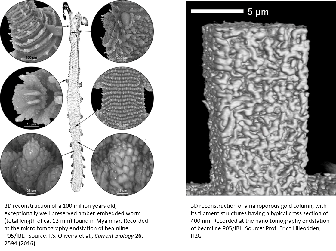
X-ray microscopy, micro tomography and nano tomography
… are state-of-the-art techniques routinely provided at the beamline P05/IBL. Within an energy range of 5 – 50 keV the application of attenuation contrast tomography delivers high resolution 3D reconstructions of complex, even strongly attenuating microstructures, with resolutions of ca. 1 µm at the micro tomography setup and down to 100 nm at the nano tomography setup.
The nano tomography setup allows a worldwide uniquely long sample-detector distance of up to 20 m and provides excellent conditions for “cone-beam” X-ray microscopy. If highest resolution is not essential, a large field of view over 7 mm is available to record micro tomography data from extended samples. Due to the wide energy range, all these techniques can be also combined with in-situ techniques, e.g. imaging data can be recorded even from within complex in situ sample environments.
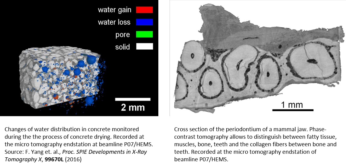
Phase contrast micro tomography
… can provide high contrast data despite low density contrast or when high photon energies are essential. This technique has been implemented at the beamlines P07/HEMS and P05/IBL, thereby throughout an energy range of 5 – 100 keV. It is ideal to visualize microstructures in samples that demand a high dynamic range in contrast with a resolution of down to 5 µm, depending on material and photon energy. Despite the high sensitivity even in low contrast samples, typical acquisition times are similar to attenuation contrast experiments and so is the large field of view of up to 7 mm. Due to the wide energy range, phase contrast imaging data can too be recorded even from within extended sample environments, providing the conditions required for in situ Experiments.
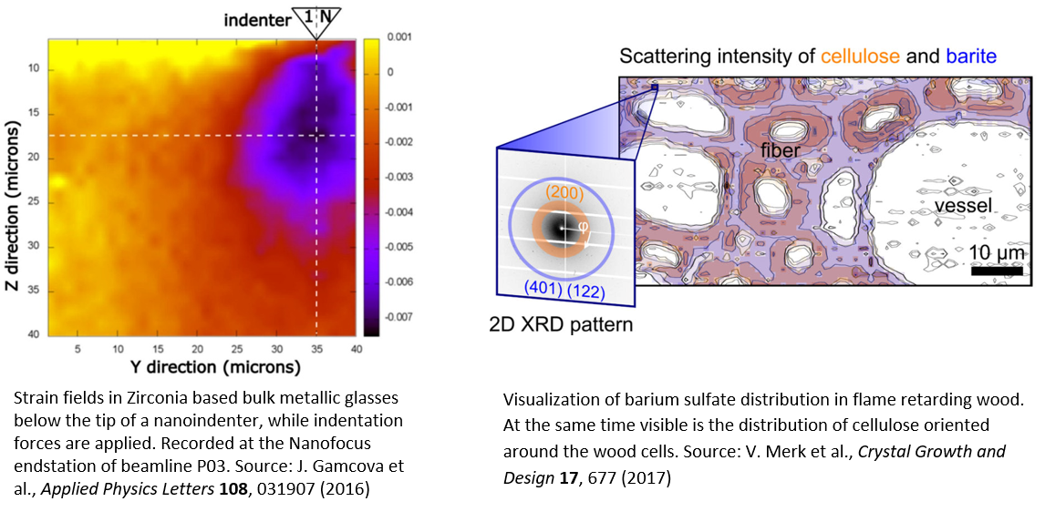
Scanning X-ray nanodiffraction
… makes use of diffraction contrast and provides atomic structural information with sub-µm resolution from crystalline and semi-crystalline materials like metals, biomaterials or synthetic compounds. It yields visual maps of residual stress profiles, crystallographic texture, phase composition, i.e. structural information that is not accessible to optical methods.
The Nanofocus Endstation of P03 beamline is a setup fully dedicated to this technique. With a beam size of 250 x 350 nm² throughout the energy range of 8 – 23 keV and a long focal distance X-ray optics with 8 cm clear focal distance it provides conditions to accommodate even extended in situ sample environments. Data can be recorded in small and wide angle geometry (SAXS and WAXS) and from within complex “in situ” sample environments, like pressure cells, tensile rigs or nanoindentation devices.
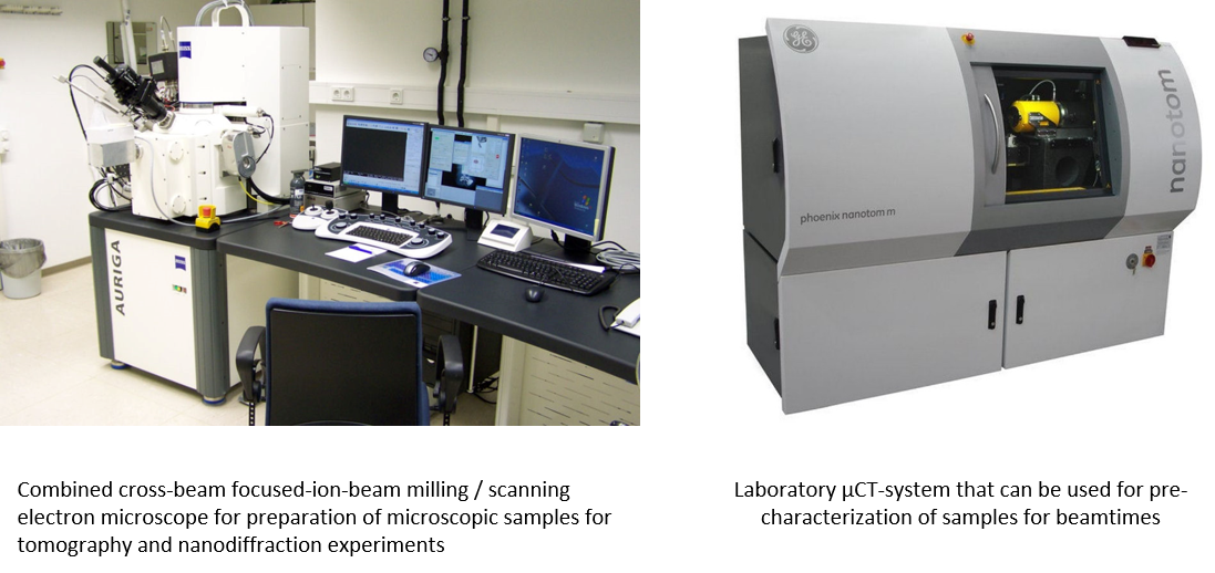
FIB/SEM and lab microtomography system
The quality of data from X-ray tomography and nanodiffraction experiments often relies on precisely manufactured samples and a comprehensive pre-characterization. For manufacturing precisely shaped microscopic samples, essential for nanotomography experiments, we use our cross-beam FIB/SEM system (Zeiss Auriga focused ion beam milling / scanning electron microscope) while our lab micro CT system (GE Nanotom) is used for obtaining 3D tomographic data outside of beam times.